If you've never experienced a cancer scare, it's easily to underestimate how challenging it can be. Even the best diagnostic tools can miss things, and you might spend weeks, months, or even years wondering if surgery, chemotherapy, or radiation really managed to eradicate all of the cancerous tissue. New innovations in magnetic resonance imaging can help put these fears to rest for doctors and patients alike.
The body has its own natural magnetic properties. The body contains abundant hydrogen atoms, which have a nucleus consisting of a single proton. Each of these protons has a north and south pole, like the Earth or a bar magnet.
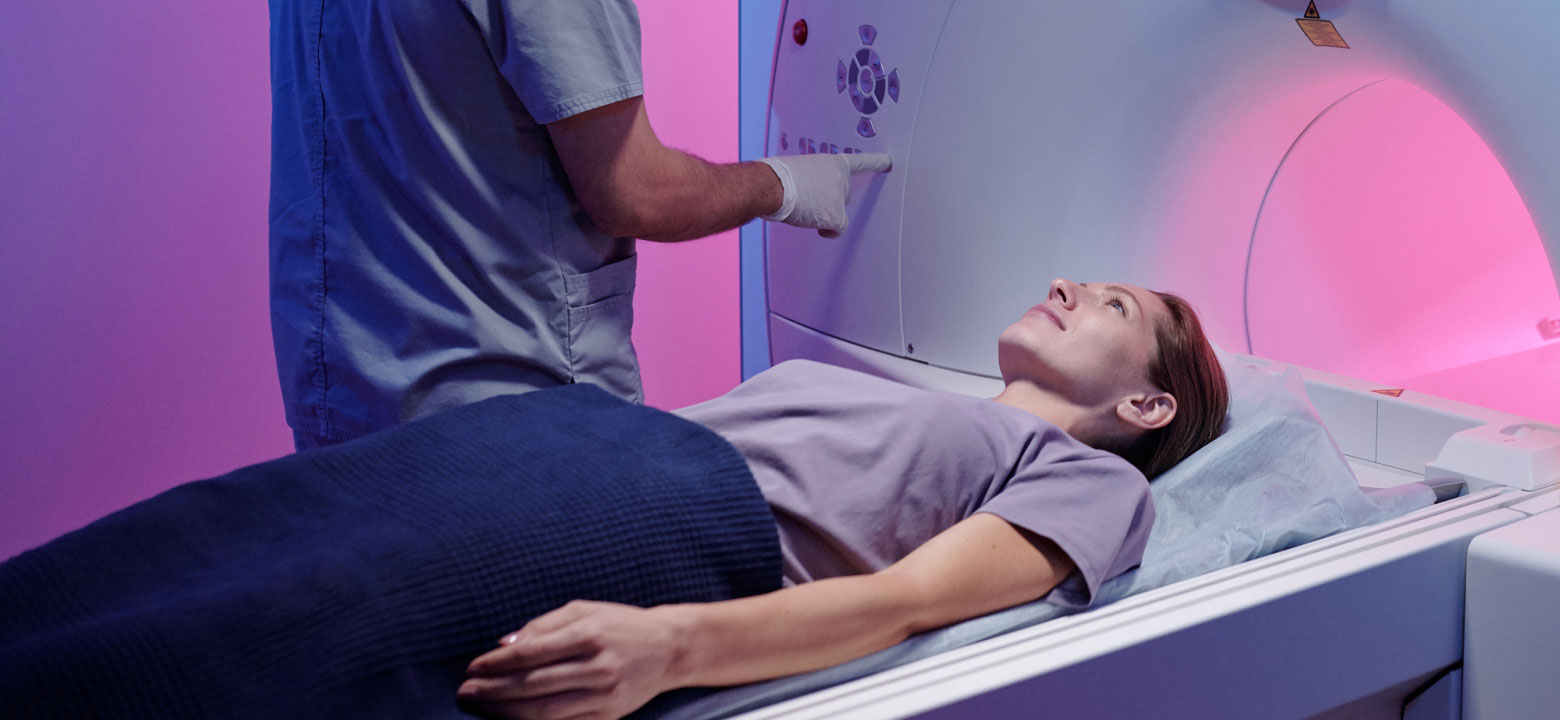
MRI machines have very powerful magnets that create a strong field. When a person is placed inside of one of these machines, all their hydrogen atoms' poles line up. When you add a radio frequency, the hydrogen atoms' nuclei resonate, and the magnetic field is deflected. Once the frequency is turned off, a signal is released that the machine uses to generate an image of the tissues inside of the body. Different tissues release signals at different rates when the frequency switches off, which is how we're able to tell the difference between bone, muscle, fat, and other structures.
Doctors and technicians can detect abnormal growths using MRI scans since abnormal tissue usually has a different water content (and therefore concentration of hydrogen atoms) than healthy tissue. Unfortunately, MRIs can have trouble differentiating between various types of diseased tissues, which makes diagnosis difficult.
With all the above in mind, researchers at the University of Waterloo developed a new technique, named synthetic correlated diffusion imaging, for using magnetic resonance to highlight cancerous tissue in a more specific fashion. This involves synthesizing and mixing MRI signals using a variety of timings and pulse strengths.
Since irregular cell growth causes differences in how water moves through cancerous tissue as opposed to regular tissue, synthetic correlated diffusion imaging is very good at indicating cancerous tissue when compared to regular tissue. Cancerous tissues actually appear to glow, which makes them stand out sharply.
So far, it's proven very helpful in the detection of prostate cancer. While prostate cancer generally has a pretty good survival rate overall, this drops off sharply in cases where there are metastases outside of the prostate. This underlines the need for early detection and diagnosis, but imaging has always been of limited use. Transrectal ultrasounds are helpful for guiding biopsies, but not good at delineating surrounding tissue from prostate tumors. Conventional MRIs can lead to misdiagnosis, as prostate cancer can be hard to differentiate from infections, inflammation, or hemorrhages.
Synthetic correlated diffusion imaging is not only able to provide strong indications of the presence of prostate cancer, it clearly delineates between healthy and cancerous tissue. It also shows promise for breast cancer and other tumors, which could provide earlier diagnoses, more peace of mind, and better outcomes for patients.
Prostate cancer is the second most common cancer in men around the world, but, until now, hasn't really had a standard method for diagnosis. Prostate-specific antigen tests often lead to overdiagnosis, while transrectal ultrasounds and MRIs can result in false positives or false negatives. Synthetic correlated diffusion imaging is a promising new method of highlighting cancer and clearly observing the margins of the disease, since it's able to delineate between abnormal cell growth and healthy tissue. In addition to providing a potential avenue for accurately diagnosing prostate cancer, it may revolutionize the imaging and diagnosis of other cancers as well.